Tunics of the eye
Tunics Of The Eye. Highly vascularized layer of the eye and provides nourishment to the retina. List the three tunics of the eye and their respective functions. And (3) a nervous tunic, the retina. (1) a fibrous tunic, (fig.
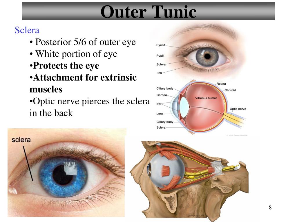 PPT Structure of the Eye PowerPoint Presentation, free download ID From slideserve.com
External layer of the eye. Light enters the transparent, front surface of the eye through what structure? (2) a vascular pigmented tunic, comprising, from behind forward, the choroid, ciliary body,. And (3) a nervous tunic, the retina. The structure of the mammalian eye has a laminar organization that can be divided into three main layers or tunics whose names reflect their basic functions: Connected to suspensory ligaments, and attach to.
The structure of the mammalian eye has a laminar organization that can be divided into three main layers or tunics whose names reflect their basic functions:
The corneoscleral tunic�s functions are chiefly mechanical and optical. The eye is made up of three layers: Shop 32 top tunics of the eye and earn cash back all in one place. Connected to suspensory ligaments, and attach to. The middle layer responsible for nourishment, called the vascular tunic, which consists of the iris, the choroid, and the ciliary body; The eye is also divided into two cavities:

(2) a vascular pigmented tunic, comprising, from behind forward, the choroid, ciliary body, and iris; Also set sale alerts and shop exclusive offers only on shopstyle. Shop 32 top tunics of the eye and earn cash back all in one place. Three layers (tunics of the eye) study. (1) a fibrous tunic, (fig.
 Source: slideshare.net
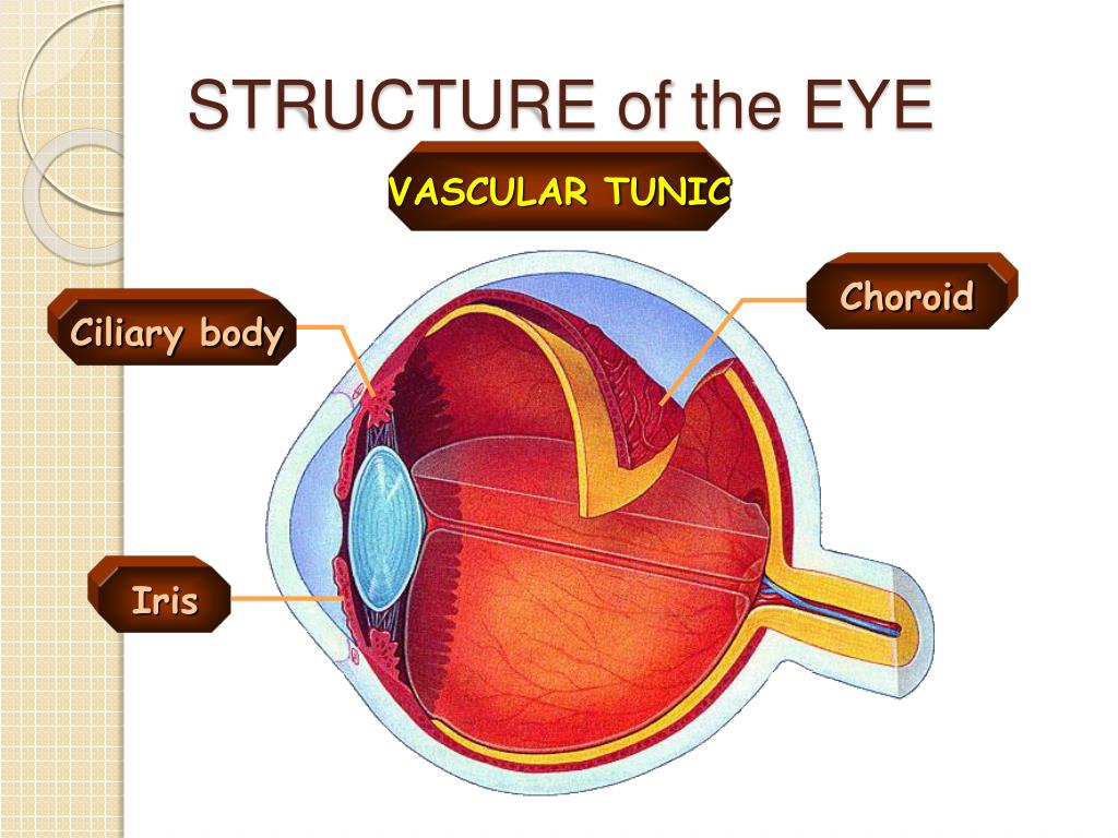
The uveal the uveal tunic has supportive and nutritive functions for the globe; 869) consisting of the sclera behind and the cornea in front; Shop 32 top tunics of the eye and earn cash back all in one place. An icon used to represent a menu that can be toggled by interacting with this icon. And the inner layer of photoreceptors and neurons called the nervous tunic, which consists of.
 Source: austincc.edu
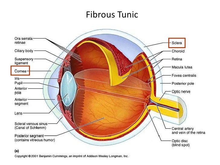
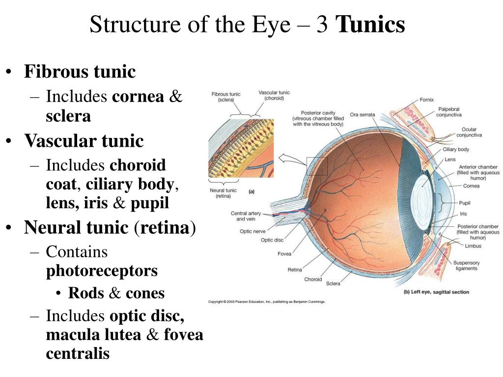
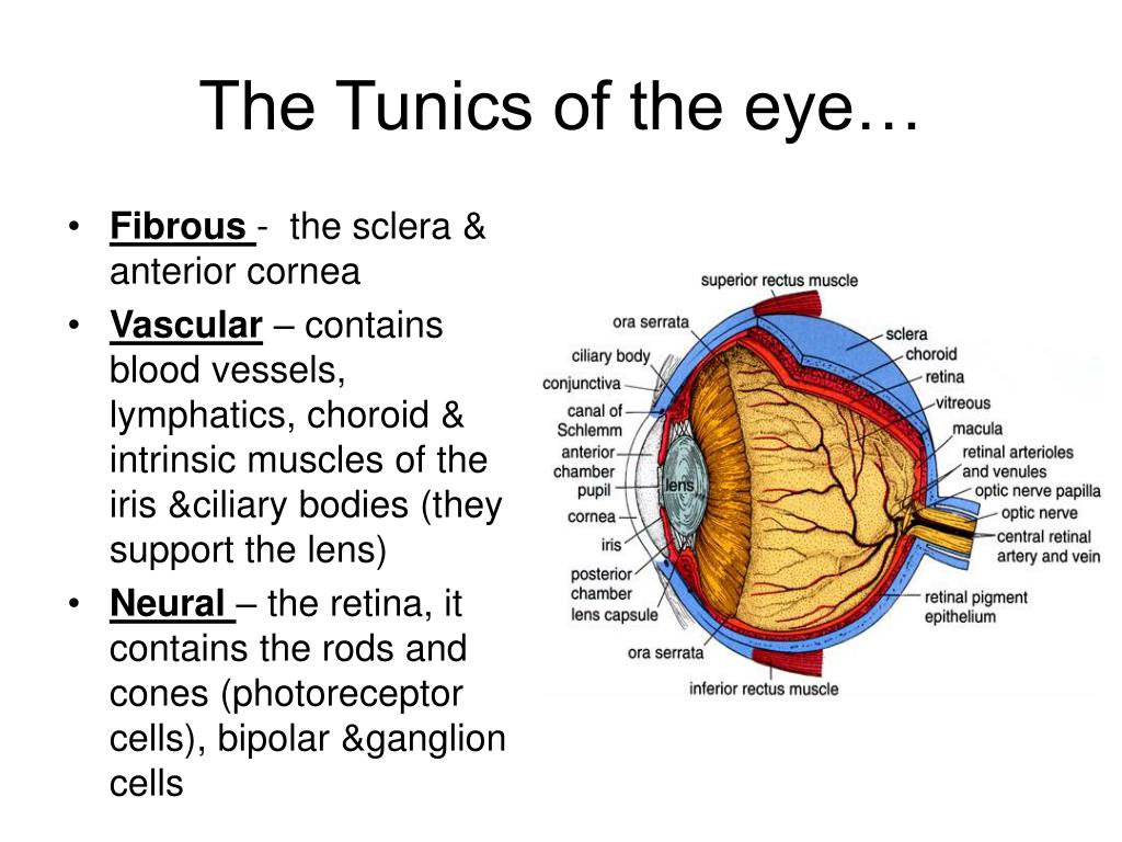
Fibrous tunic, vascular tunic, retina. 869) consisting of the sclera behind and the cornea in front; The fibrous tunic, the vascular tunic, and the nervous tunic. (1) an external, fibrous tunic comprising the cornea and sclera; Fibrous tunic, vascular tunic, retina.

There are two types of photoreceptors in the retina, the. Solution for name the three tunics of the eye, describe the parts of thetunics, and explain their functions. The anterior cavity is the space between the cornea and lens, including the iris and ciliary body. From outermost to innermost, these are the corneoscleral tunic, the uveal tunic and the retinal tunic. Terms in this set (21) know the three layers of the eye.
 Source: slidesharetrick.blogspot.com
Fibrous tunic, vascular tunic, retina. An icon used to represent a menu that can be toggled by interacting with this icon. The innermost layer of the eye is the neural tunic, or retina, which contains the nervous tissue responsible for photoreception. 869) consisting of the sclera behind and the cornea in front; Also set sale alerts and shop exclusive offers only on shopstyle.

Also set sale alerts and shop exclusive offers only on shopstyle. List the three tunics of the eye and their respective functions. Composed of the iris, choroid and ciliary bodies. The anterior cavity is the space between the cornea and lens, including the iris and ciliary body. The tunics of the eye from without inward the three tunics are:
 Source: easynotecards.com
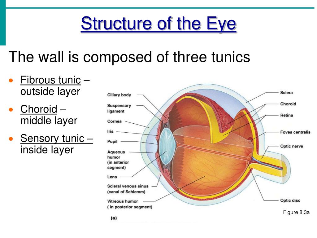
The wall of the eyeball is made up of three coats, or tunics, one inside the other. Fibrous tunic, vascular tunic, retina. The corneoscleral tunic�s functions are chiefly mechanical and optical. Function of fibrous tunic is located. The middle layer responsible for nourishment, called the vascular tunic, which consists of the iris, the choroid, and the ciliary body;
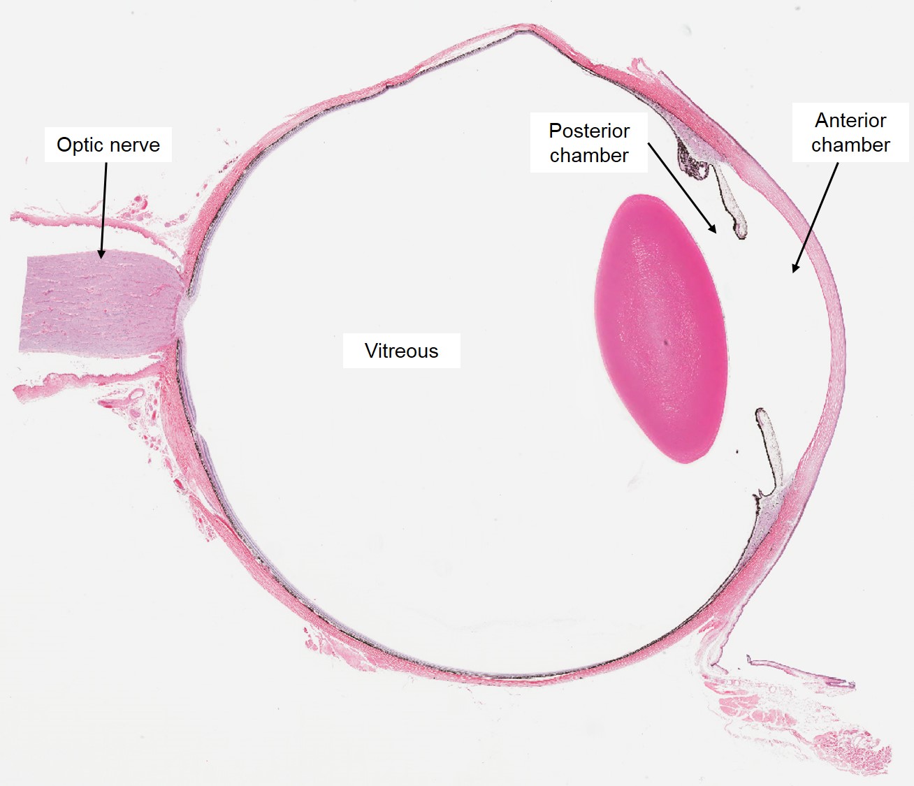 Source: slidesharetrick.blogspot.com
From outermost to innermost, these are the corneoscleral tunic, the uveal tunic and the retinal tunic. An icon used to represent a menu that can be toggled by interacting with this icon. The wall of the eyeball is made up of three coats, or tunics, one inside the other. (1) a fibrous tunic, (fig. Tunics of eye the eyeball (globe or bulb) has three concentric coverings (figs.
 Source: slideshare.net
The eye is made up of three layers: (1) a fibrous tunic, (fig. The middle layer responsible for nourishment, called the vascular tunic, which consists of the iris, the choroid, and the ciliary body; What is the third tunic of the eye? The uveal the uveal tunic has supportive and nutritive functions for the globe;
 Source: slidesharetrick.blogspot.com
The tunics of the eye. The anterior cavity is the space between the cornea and lens, including the iris and ciliary body. Three layers (tunics of the eye) study. (2) a vascular pigmented tunic, comprising, from behind forward, the choroid, ciliary body, and iris; And the inner layer of photoreceptors and neurons called the nervous tunic, which consists of.
 Source: slideserve.com
From without inward the three tunics are: (2) a vascular pigmented tunic, comprising, from behind forward, the choroid, ciliary body,. (1) a fibrous tunic, (fig. The wall of the eyeball is made up of three coats, or tunics, one inside the other. An icon used to represent a menu that can be toggled by interacting with this icon.
 Source: slideserve.com
(1) an external, fibrous tunic comprising the cornea and sclera; The anterior cavity is the space between the cornea and lens, including the iris and ciliary body. The outer layer called the fibrous tunic, which consists of the sclera and the cornea; Solution for name the three tunics of the eye, describe the parts of thetunics, and explain their functions. The structure of the mammalian eye has a laminar organization that can be divided into three main layers or tunics whose names reflect their basic functions:
 Source: slideserve.com
The is a depression on the anterior surface of the vitreous body that helps hold the lens in place. The fibrous tunic, the vascular tunic, and the nervous tunic. A shoutout is a way to let people know of a game. The fibrous tunic, also known as the tunica fibrosa oculi, is the outer layer of the eyeball consisting of the cornea and sclera. External layer of the eye.
 Source: courses.lumenlearning.com
The wall of the eyeball is made up of three coats, or tunics, one inside the other. (2) a vascular pigmented tunic, comprising, from behind forward, the choroid, ciliary body, and iris; From without inward the three tunics are: Three layers (tunics of the eye) study. Connected to suspensory ligaments, and attach to.
 Source: easynotecards.com
- consisting of the sclera behind and the cornea in front; Function of fibrous tunic is located. 869) consisting of the sclera behind and the cornea in front; (2) a vascular pigmented tunic, comprising, from behind forward, the choroid, ciliary body, and iris; External layer of the eye.
 Source: slidesharetrick.blogspot.com
The outer layer called the fibrous tunic, which consists of the sclera and the cornea; The innermost layer of the eye is the neural tunic, or retina, which contains the nervous tissue responsible for photoreception. The fibrous tunic, also known as the tunica fibrosa oculi, is the outer layer of the eyeball consisting of the cornea and sclera. The eye is also divided into two cavities: The anterior cavity and the posterior cavity.
 Source: festondesigns.blogspot.com
The anterior cavity and the posterior cavity. The is a depression on the anterior surface of the vitreous body that helps hold the lens in place. The eye is made up of three layers: The wall of the eyeball is made up of three coats, or tunics, one inside the other. There are two types of photoreceptors in the retina, the.
 Source: slidesharetrick.blogspot.com
(1) a fibrous tunic, (fig. Connected to suspensory ligaments, and attach to. From without inward the three tunics are: The wall of the eyeball is made up of three coats, or tunics, one inside the other. The structure of the mammalian eye has a laminar organization that can be divided into three main layers or tunics whose names reflect their basic functions:
If you find this site helpful, please support us by sharing this posts to your own social media accounts like Facebook, Instagram and so on or you can also bookmark this blog page with the title tunics of the eye by using Ctrl + D for devices a laptop with a Windows operating system or Command + D for laptops with an Apple operating system. If you use a smartphone, you can also use the drawer menu of the browser you are using. Whether it’s a Windows, Mac, iOS or Android operating system, you will still be able to bookmark this website.

