X ray positioning pictures
X Ray Positioning Pictures. Central ray is perpendicular to the third metacarpel joint. Position of patient lying on the side (left or right) with a vertical beam angled at 15 degrees. The patient is supine (on an inclined radiographic table) with the head lower than the feet. The patient is seated alongside the table;
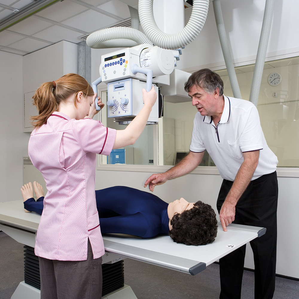
 Simulated setup showing positioning of the portable Xray unit for From researchgate.net
This provides a clear view of the open joint spaces and soft tissues around the knee joint as well. Fully extended arm and forearm, in a pronated position ensure the anterior portion of the elbow is roughly 45 degrees from the ir technical factors. The smaller image indicates positioning for frontal bone and maxilla. Anterior is towards the front of the body latin. Central ray is perpendicular with the 3rd metacarpel joint. Position of part remove dentures, facial jewelry, earrings, and anything from the hair.
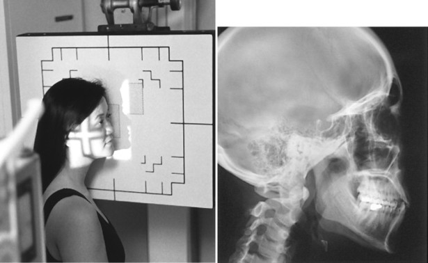
The smaller image indicates positioning for frontal bone and maxilla.
Central ray is perpendicular with the 3rd metacarpel joint. Superior to the distal third of the humerus It discusses radiographic positioning of the knee in pa projections. Purpose and structures shown to demonstrate a posteroanterior (pa) image of the knee. Central ray is perpendicular to the third metacarpel joint. Stair step sponge is used for support and to help maintain position.
 Source: polymedlab.ph
Stair step sponge is used for support and to help maintain position. Superior to the distal third of the humerus The patient is supine (on an inclined radiographic table) with the head lower than the feet. The patient is positioned supine on the radiographic table, with arms placed at the. Elbow is 90 deg, arm fully supported.used when not using the stair step sponge, from the lateral position, the hand forms a 45 deg angle with the ir.
 Source: radiologykey.com
See more ideas about radiography, radiology, radiologic technology. The smaller image indicates positioning for frontal bone and maxilla. The patient is seated alongside the table; The patient is supine (on an inclined radiographic table) with the head lower than the feet. See more ideas about radiography, radiology, x ray.
 Source: adam-rouilly.co.uk
After the remote couch correction, the treatment is immediately started. After the remote couch correction, the treatment is immediately started. The smaller image indicates positioning for frontal bone and maxilla. Positions are learned by the radiographer according to body part in. Position of part remove dentures, facial jewelry, earrings, and anything from the hair.
 Source: fluororadpro.com
Purpose and structures shown an additional view to evaluate the mandible. This provides a clear view of the open joint spaces and soft tissues around the knee joint as well. The patient is seated alongside the table; Right side touches the cassette. The smaller image indicates positioning for frontal bone and maxilla.
 Source: worldpoint.com
Purpose and structures shown to demonstrate a posteroanterior (pa) image of the knee. Position of part remove dentures, facial jewelry, earrings, and anything from the hair. The center of the cassette should be midway between the asis and the pubic symphysis. Superior to the distal third of the humerus The patient is seated alongside the table;
 Source: researchgate.net
It discusses radiographic positioning of the knee in pa projections. Purpose and structures shown an additional view to evaluate the mandible. The patient is seated alongside the table; Positions are learned by the radiographer according to body part in. Position of patient lying on the side (left or right) with a vertical beam angled at 15 degrees.
 Source: radiologykey.com
This provides a clear view of the open joint spaces and soft tissues around the knee joint as well. Position of part remove dentures, facial jewelry, earrings, and anything from the hair. See more ideas about radiography, radiology, x ray. The patient is seated alongside the table; The center of the cassette should be midway between the asis and the pubic symphysis.
 Source: radiopaedia.org
Superior to the distal third of the humerus See more ideas about radiography, radiology, radiologic technology. Purpose and structures shown an additional view to evaluate the mandible. Fully extended arm and forearm, in a pronated position ensure the anterior portion of the elbow is roughly 45 degrees from the ir technical factors. The center of the cassette should be midway between the asis and the pubic symphysis.
 Source: sciencephoto.com
Position of patient lying on the side (left or right) with a vertical beam angled at 15 degrees. Elbow is 90 deg, arm fully supported.used when not using the stair step sponge, from the lateral position, the hand forms a 45 deg angle with the ir. The smaller image indicates positioning for frontal bone and maxilla. Central ray is perpendicular to the third metacarpel joint. The patient is positioned supine on the radiographic table, with arms placed at the.
 Source: radiopaedia.org
The center of the cassette should be midway between the asis and the pubic symphysis. The smaller image indicates positioning for frontal bone and maxilla. It discusses radiographic positioning of the knee in pa projections. The patient is seated alongside the table; Position of part remove dentures, facial jewelry, earrings, and anything from the hair.
Source: vetpol.co.uk
Superior to the distal third of the humerus Central ray is perpendicular with the 3rd metacarpel joint. Stair step sponge is used for support and to help maintain position. Position of part remove dentures, facial jewelry, earrings, and anything from the hair. The smaller image indicates positioning for frontal bone and maxilla.
 Source: youtube.com
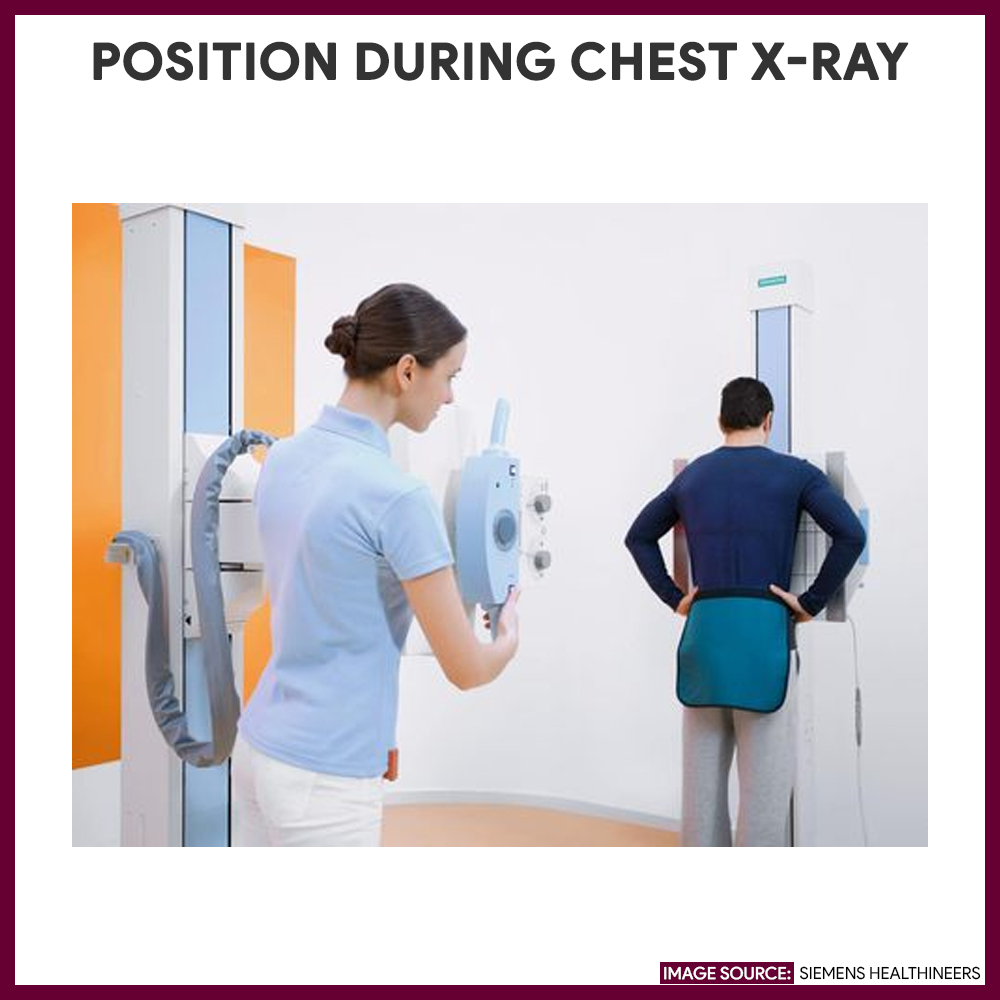
Right side touches the cassette. Right side touches the cassette. After the remote couch correction, the treatment is immediately started. See more ideas about radiography, radiology, x ray. It discusses radiographic positioning of the knee in pa projections.
 Source: nurseslabs.com
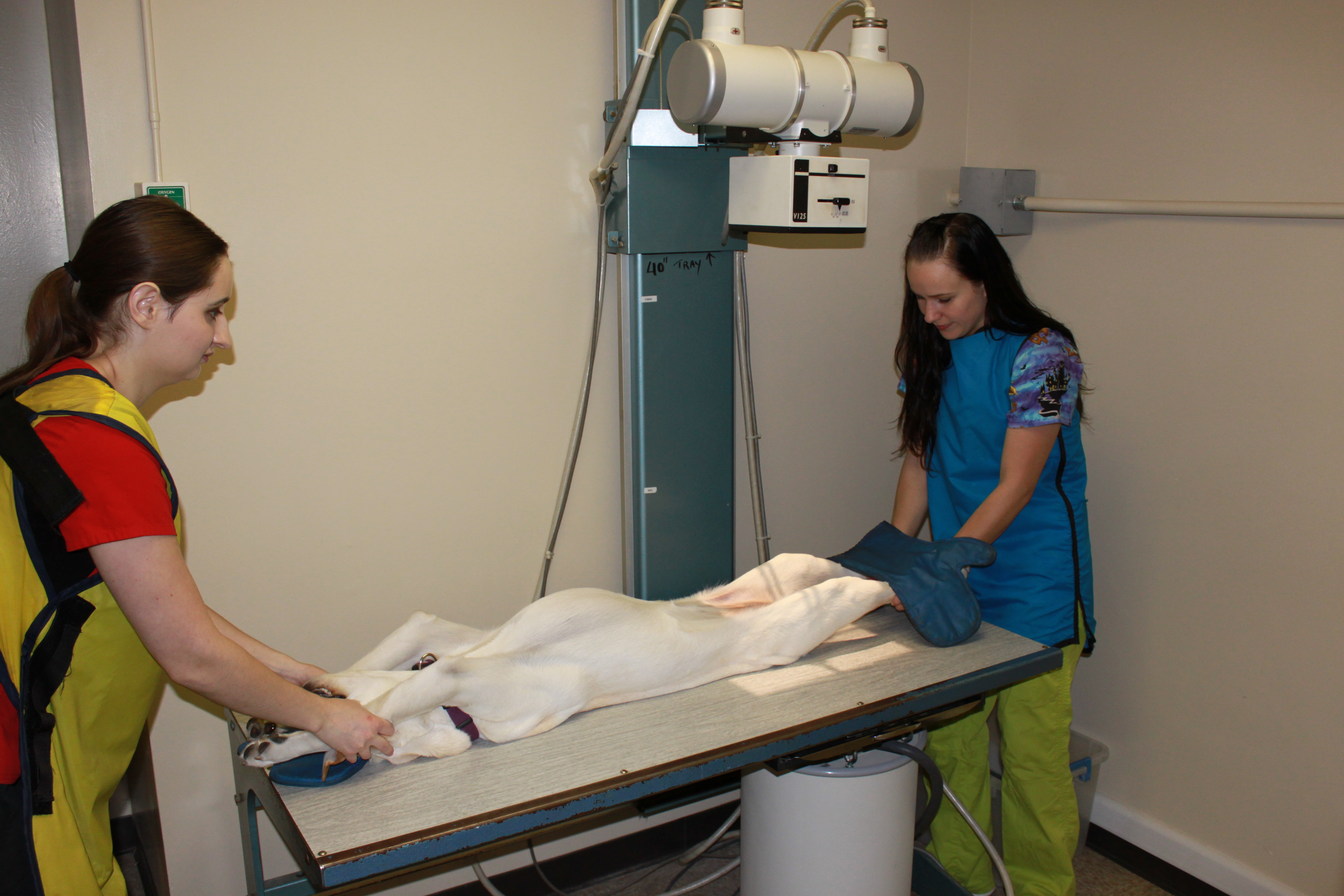
See more ideas about radiography, radiology, x ray. The center of the cassette should be midway between the asis and the pubic symphysis. Stair step sponge is used for support and to help maintain position. The patient is supine (on an inclined radiographic table) with the head lower than the feet. This provides a clear view of the open joint spaces and soft tissues around the knee joint as well.
 Source: riverviewanimalclinicclarkston.com
Right side touches the cassette. Purpose and structures shown an additional view to evaluate the mandible. Right side touches the cassette. The patient is supine (on an inclined radiographic table) with the head lower than the feet. Central ray is perpendicular with the 3rd metacarpel joint.
 Source: fluororadpro.com
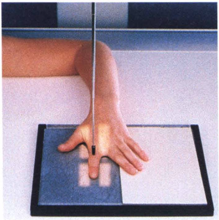
Positions are learned by the radiographer according to body part in. See more ideas about radiography, radiology, radiologic technology. Stair step sponge is used for support and to help maintain position. The patient is positioned supine on the radiographic table, with arms placed at the. Position of part remove dentures, facial jewelry, earrings, and anything from the hair.
 Source: radtexts.blogspot.com
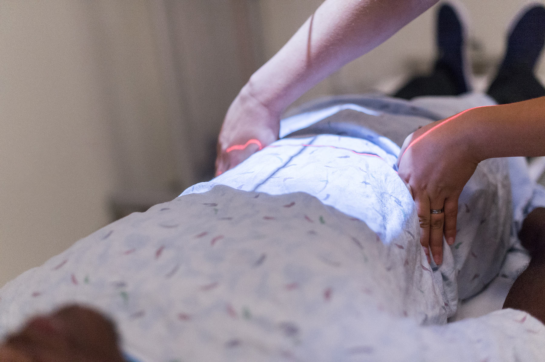
The center of the cassette should be midway between the asis and the pubic symphysis. It discusses radiographic positioning of the knee in pa projections. See more ideas about radiography, radiology, x ray. The center of the cassette should be midway between the asis and the pubic symphysis. Purpose and structures shown an additional view to evaluate the mandible.
 Source: blog.radiology.virginia.edu
Elbow is 90 deg, arm fully supported.used when not using the stair step sponge, from the lateral position, the hand forms a 45 deg angle with the ir. Central ray is perpendicular with the 3rd metacarpel joint. The center of the cassette should be midway between the asis and the pubic symphysis. The patient is supine (on an inclined radiographic table) with the head lower than the feet. Elbow is 90 deg, arm fully supported.used when not using the stair step sponge, from the lateral position, the hand forms a 45 deg angle with the ir.
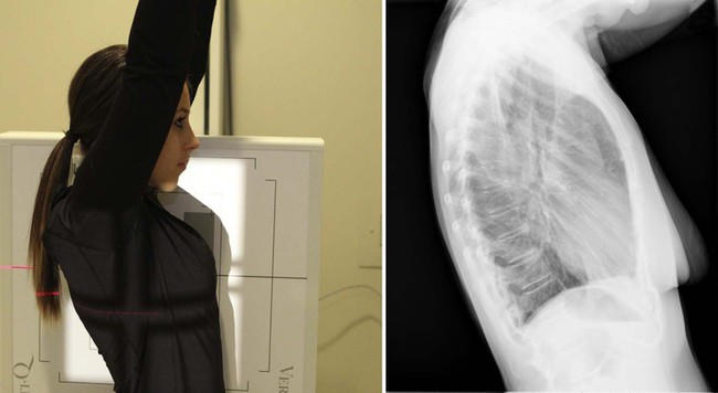 Source: radiologykey.com
Purpose and structures shown an additional view to evaluate the mandible. Central ray is perpendicular with the 3rd metacarpel joint. See more ideas about radiography, radiology, x ray. Central ray is perpendicular to the third metacarpel joint. Anterior is towards the front of the body latin.
If you find this site beneficial, please support us by sharing this posts to your own social media accounts like Facebook, Instagram and so on or you can also save this blog page with the title x ray positioning pictures by using Ctrl + D for devices a laptop with a Windows operating system or Command + D for laptops with an Apple operating system. If you use a smartphone, you can also use the drawer menu of the browser you are using. Whether it’s a Windows, Mac, iOS or Android operating system, you will still be able to bookmark this website.

