Split lung function test
Split Lung Function Test. Download scientific diagram | paired split lung function using q pet/ct and the recommended methods, i.e. A significant difference between the. In addition, a diffusing capacity test measures how easily oxygen enters the bloodstream. On the other hand, in some patients the.
 Exercise testing basic knowledge From slideshare.net
They measure lung volumes, lung capacity, rates of flow of gases, and the efficiency of gas exchange. If you’re regularly exposed to certain substances in. Lung diffusion is your ability to pass oxygen into the blood from the air sacs of the lungs, and pass carbon dioxide (co2) back into the lungs from. It is influenced by airway resistance, respiratory muscles, compliance of the lungs and/or chest wall, and ventilatory control mechanisms. This split lung perfusion (slp) combines the advantages of isolated whole lung perfusion studies with the advantages derived from the use of paired organs. On the other hand, in some patients the.
From the 133xe inhalation scan, a split lung capacity (right to left, upper, middle and lower) and t1/2 (time.
Lung volume testing is another commonly performed lung function test. 133xe inhalation scan and ordinary lung function testing were performed three times in 34 patients undergoing pulmonary resection: The study is combined with pulmonary lung function test ; Maximal voluntary ventilation (mvv) is the volume of air exhaled in 12 seconds during rapid forced breathing. The ratio between these two numbers, the fev1/fvc, tells you whether or not the patient is obstructed. “lung function tests.” american lung association:
 Source: slideserve.com
“emphysema,” “lung function tests,” “pulse oximetry.” johns hopkins medicine: They measure lung volumes, lung capacity, rates of flow of gases, and the efficiency of gas exchange. Pfts are performed by outpatient procedures called spirometry and sometimes an added test called plethysmography. From the 133xe inhalation scan, a split lung capacity (right to left, upper, middle and lower) and t1/2 (time. Using statistical tests to analyze the effects of acute.
 Source: slideshare.net
It is more precise than spirometry and measures the volume of air in the lungs, including the air that remains at the end of a normal breath. For some of the test measurements, the client can breathe normally and quietly. Doctors carry out pulmonary function tests to check how well a person’s lungs work and identify any issues. They measure lung volumes, lung capacity, rates of flow of gases, and the efficiency of gas exchange. Pfts are performed by outpatient procedures called spirometry and sometimes an added test called plethysmography.
 Source: researchgate.net
On the other hand, in some patients the. Pulmonary function tests (pfts) are noninvasive tests that show how well the lungs are working. 133xe inhalation scan and ordinary lung function testing were performed three times in 34 patients undergoing pulmonary resection: If the fev1/fvc is too low, the patient is obstructed and if the fev1/fvc is either normal or higher than normal, then the patient is not obstructed. The tests measure lung volume, capacity, rates of flow, and gas exchange.
 Source: pmj.bmj.com
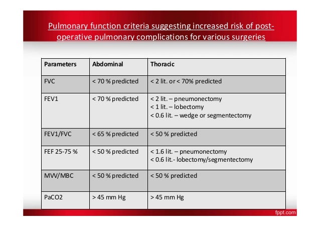
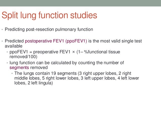
If the patient does not meet the above criteria on routine pft, and if the fev1 volume is less than 2 liter, we need to perform split lung function testing. Other tests require forced inhalation or exhalation after a deep breath. They measure lung volumes, lung capacity, rates of flow of gases, and the efficiency of gas exchange. Exercise testing helps evaluate causes of. There are 2 definitions of a “normal” fev1/fvc that are commonly used.
 Source: jtcvs.org
Pulmonary function tests (pfts) are a group of noninvasive tests that measure how well your lungs work. Exercise testing helps evaluate causes of. Pulmonary function tests are used to assess how well your lungs are functioning. The study is combined with pulmonary lung function test ; If the fev1/fvc is too low, the patient is obstructed and if the fev1/fvc is either normal or higher than normal, then the patient is not obstructed.
 Source: slideshare.net
This information can help your healthcare provider diagnose and decide the treatment of certain lung disorders. Dlco, otherwise known as a gas diffusion test, looks at how well the lungs actually absorb oxygen. Pulmonary function tests are used to assess how well your lungs are functioning. It is more precise than spirometry and measures the volume of air in the lungs, including the air that remains at the end of a normal breath. If the patient does not meet the above criteria on routine pft, and if the fev1 volume is less than 2 liter, we need to perform split lung function testing.
 Source: slideshare.net
The mvv is the total volume of air exhaled during 12 seconds of rapid, deep breathing, which can be compared with a predicted mvv defined as the forced expiratory volume in 1 second (fev1) × 35 or 40. Mvv tests the overall function of the respiratory system. This split lung perfusion (slp) combines the advantages of isolated whole lung perfusion studies with the advantages derived from the use of paired organs. The tests measure lung volume, capacity, rates of flow, and gas exchange. The ratio between these two numbers, the fev1/fvc, tells you whether or not the patient is obstructed.
 Source: slideserve.com
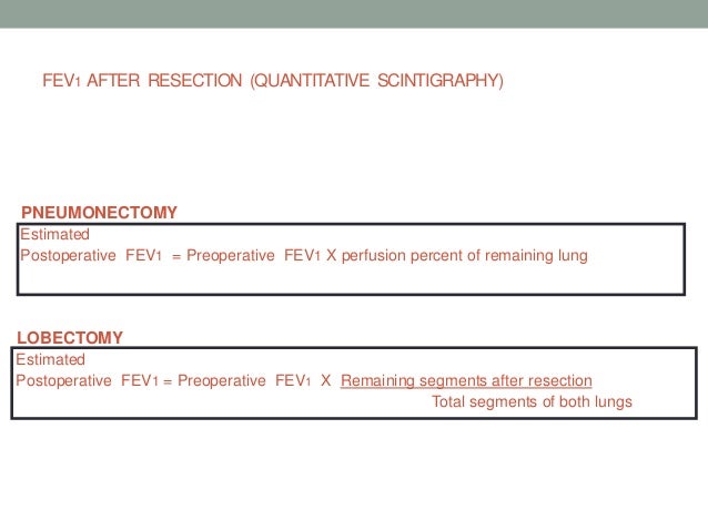
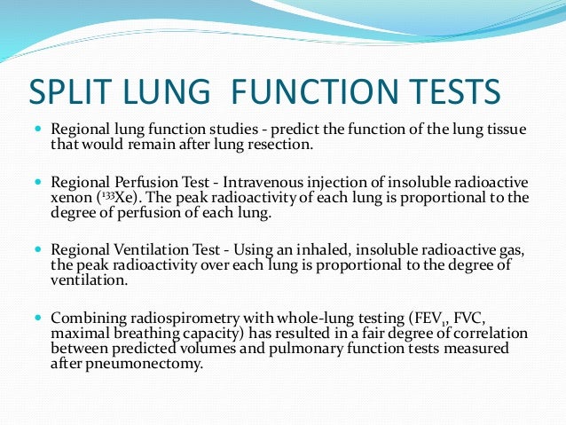
The maximal voluntary ventilation (mvv) is another measure of the neuromuscular and respiratory systems. From the 133xe inhalation scan, a split lung capacity (right to left, upper, middle and lower) and t1/2 (time. Dlco, otherwise known as a gas diffusion test, looks at how well the lungs actually absorb oxygen. The study is combined with pulmonary lung function test ; Regional perfusion tests (using iv 133xe) allow one to examine the relative perfusion of each lung.
 Source: slideshare.net
Sometimes, they will be asked to inhale a different gas or a medicine to see how it changes test results. This split lung perfusion (slp) combines the advantages of isolated whole lung perfusion studies with the advantages derived from the use of paired organs. Pulmonary function tests are used to assess how well your lungs are functioning. Regional perfusion tests (using iv 133xe) allow one to examine the relative perfusion of each lung. It is more precise than spirometry and measures the volume of air in the lungs, including the air that remains at the end of a normal breath.
 Source: slideshare.net
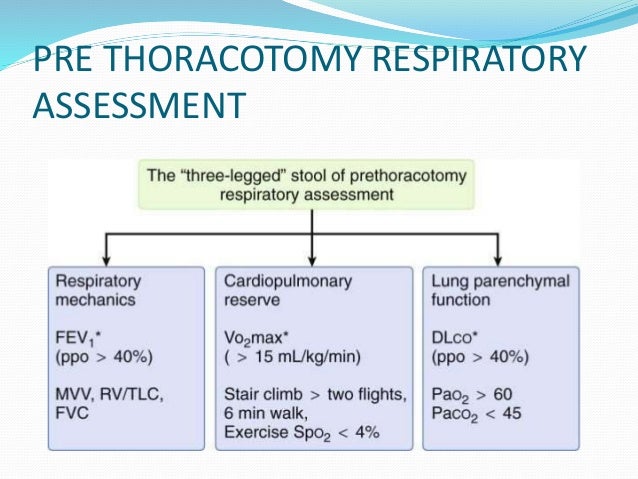
The mvv is the total volume of air exhaled during 12 seconds of rapid, deep breathing, which can be compared with a predicted mvv defined as the forced expiratory volume in 1 second (fev1) × 35 or 40. Exercise testing helps evaluate causes of. Regional perfusion tests (using iv 133xe) allow one to examine the relative perfusion of each lung. Among lung function measures, fev1 and dlco are the most useful predictors of postoperative outcomes. Mvv tests the overall function of the respiratory system.
 Source: researchgate.net
There are 2 types of disorders that cause problems with air. In addition, a diffusing capacity test measures how easily oxygen enters the bloodstream. Before surgery, and one and six months postoperatively. The mvv is the total volume of air exhaled during 12 seconds of rapid, deep breathing, which can be compared with a predicted mvv defined as the forced expiratory volume in 1 second (fev1) × 35 or 40. The ratio between these two numbers, the fev1/fvc, tells you whether or not the patient is obstructed.
 Source: slideshare.net
There are 2 types of disorders that cause problems with air. There are 2 definitions of a “normal” fev1/fvc that are commonly used. The ratio between these two numbers, the fev1/fvc, tells you whether or not the patient is obstructed. This measures the split lung function using the ct attenuation density instead of radionuclide signal intensity. Before surgery, and one and six months postoperatively.
 Source: slideshare.net
Regional perfusion tests (using iv 133xe) allow one to examine the relative perfusion of each lung. They measure lung volumes, lung capacity, rates of flow of gases, and the efficiency of gas exchange. “lung function tests.” american lung association: This measures the split lung function using the ct attenuation density instead of radionuclide signal intensity. Exercise testing helps evaluate causes of.
 Source: slideserve.com
If you’re regularly exposed to certain substances in. Lung volume testing is another commonly performed lung function test. Lungs with tumor may not be contributing to total fev1 volume and thus removal of it may not significantly affect pulmonary function. Mvv tests the overall function of the respiratory system. Lung diffusion is your ability to pass oxygen into the blood from the air sacs of the lungs, and pass carbon dioxide (co2) back into the lungs from.
 Source: slideshare.net
Pulmonary function tests, or pfts, measure how well the lungs work. Regional perfusion tests (using iv 133xe) allow one to examine the relative perfusion of each lung. From the 133xe inhalation scan, a split lung capacity (right to left, upper, middle and lower) and t1/2 (time. Pulmonary function tests, or pfts, measure how well the lungs work. This measures the split lung function using the ct attenuation density instead of radionuclide signal intensity.
 Source: thoracickey.com
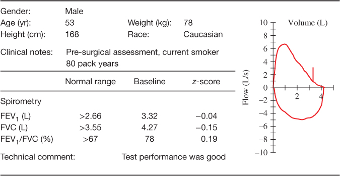
The ratio between these two numbers, the fev1/fvc, tells you whether or not the patient is obstructed. If the fev1/fvc is too low, the patient is obstructed and if the fev1/fvc is either normal or higher than normal, then the patient is not obstructed. The study is combined with pulmonary lung function test ; Pulmonary function tests, or pfts, measure how well the lungs work. A significant difference between the.
 Source: anaesthesiajournal.co.uk
During that breath hold time, you’re inhaling a. Other tests require forced inhalation or exhalation after a deep breath. On the other hand, in some patients the. Pfts are performed by outpatient procedures called spirometry and sometimes an added test called plethysmography. Lungs with tumor may not be contributing to total fev1 volume and thus removal of it may not significantly affect pulmonary function.
 Source: researchgate.net
It is influenced by airway resistance, respiratory muscles, compliance of the lungs and/or chest wall, and ventilatory control mechanisms. On the other hand, in some patients the. Lungs with tumor may not be contributing to total fev1 volume and thus removal of it may not significantly affect pulmonary function. Pulmonary function tests, or pfts, measure how well the lungs work. This measures the split lung function using the ct attenuation density instead of radionuclide signal intensity.
If you find this site helpful, please support us by sharing this posts to your own social media accounts like Facebook, Instagram and so on or you can also bookmark this blog page with the title split lung function test by using Ctrl + D for devices a laptop with a Windows operating system or Command + D for laptops with an Apple operating system. If you use a smartphone, you can also use the drawer menu of the browser you are using. Whether it’s a Windows, Mac, iOS or Android operating system, you will still be able to bookmark this website.


