Sinuses x ray positioning
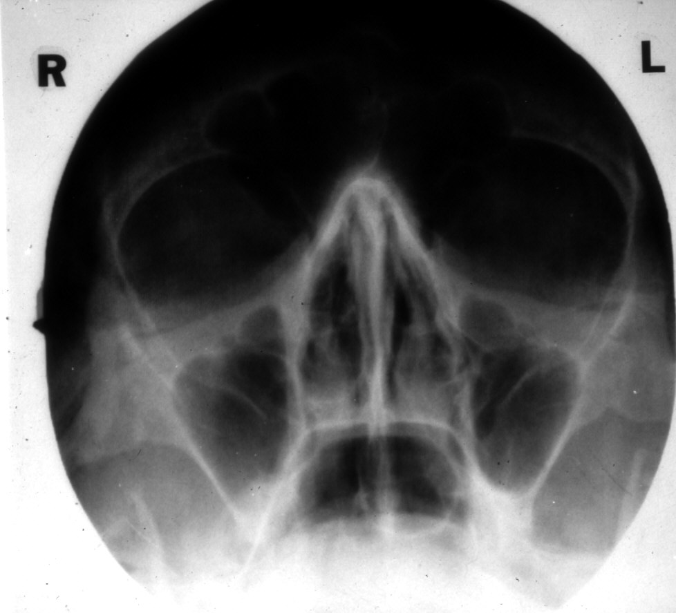
Sinuses X Ray Positioning. The overlying petrosal bone masks the maxillary sinus.by the way, you can see the superior orbital fissure and the round foramen beautifully on this projection. Frontal sinuses projected above the frontonasal suture, anterior ethmoid air cells visualised lateral to each nasal bone directly below the frontal sinuses. The caldwell (posteroanterior), waters (occipitomental), and lateral views (figs. The patient’s head should be tilted by 15 degrees.
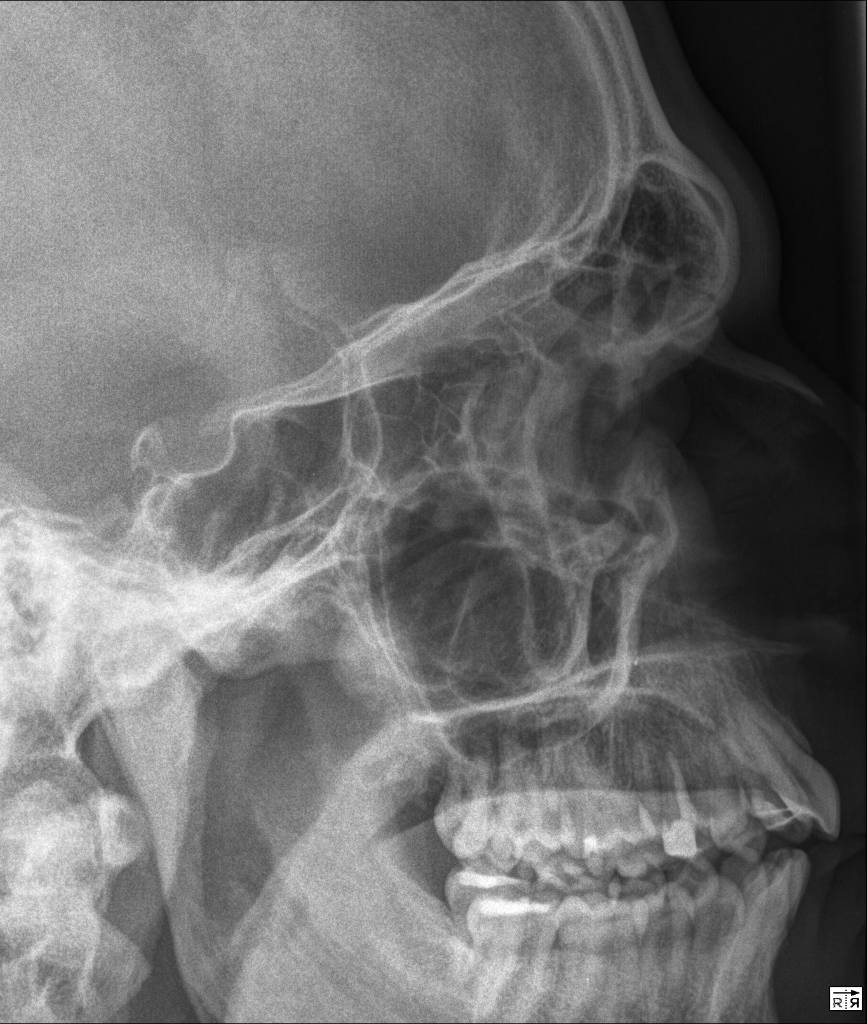 PARANASAL SINUSES LATERAL PROJECTION From buyxraysonline.com
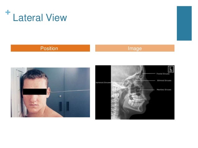
This view aids in visualizing the paranasal sinuses, especially the frontal sinus.it can help to assess 4 inflammatory conditions such as sinusitis and secondary osteomyelitis, and sinus polyps or cysts. About press copyright contact us creators advertise developers terms privacy policy & safety how youtube works test new features press copyright contact us creators. Radt 210 radiographic positioning iiisan diego mesa collegeradiographic positioning of the paranasal sinuses Sometimes additional stacking is applied, allowing to examine defects in more detail. For each setup in the tables, there is a picture demonstrating. Patient erect where possible to demonstrate fluid levels.
The image depicts the frontal, ethmoidal, and sphenoid sinuses.
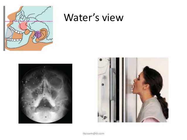
For each setup in the tables, there is a picture demonstrating. Patient positioning for skull radiography. Patient positioned same as waters view, but with mouth opened. Frontal sinuses projected above the frontonasal suture, anterior ethmoid air cells visualised lateral to each nasal bone directly below the frontal sinuses. Is recommended for evaluation of paranasal sinuses. The image depicts the frontal, ethmoidal, and sphenoid sinuses.
 Source: pinterest.jp
About press copyright contact us creators advertise developers terms privacy policy & safety how youtube works test new features press copyright contact us creators. An opaque sinus may be caused by the thickening of the bony walls, small asymmetric antra, or improper centering and rotation of the head. The image depicts the frontal, ethmoidal, and sphenoid sinuses. Patients can be imaged either erect or recumbent. Sinusitis, secondary osteomyelitis and sinus polyps.
 Source: buyxraysonline.com
This view must demonstrate sphenoid sinus through the open mouth (patient�s mouth is placed on table). The caldwell (posteroanterior), waters (occipitomental), and lateral views (figs. The overlying petrosal bone masks the maxillary sinus.by the way, you can see the superior orbital fissure and the round foramen beautifully on this projection. Other sinuses related problems such as deviated septum. The patient’s head should be tilted by 15 degrees.
 Source: youtube.com
Based on the acr appropriateness criteria ©, ct sinus. The caldwell (posteroanterior), waters (occipitomental), and lateral views (figs. Frontal sinuses projected above the frontonasal suture, anterior ethmoid air cells visualised lateral to each nasal bone directly below the frontal sinuses. Is recommended for evaluation of paranasal sinuses. Position of part remove dentures, facial jewelry, earrings, and anything from the hair.
 Source: pinterest.com
The epithelial surface of the sinuses is an extension of the nasal mucosa. Position of part remove dentures, facial jewelry, earrings, and anything from the hair. An opaque sinus may be caused by the thickening of the bony walls, small asymmetric antra, or improper centering and rotation of the head. The image depicts the frontal, ethmoidal, and sphenoid sinuses. Additionally, any fractures to the orbit may also be determined through this view.
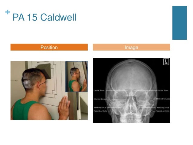 Source: slideshare.net
Paranasal sinuses water’s view caldwell view lateral view submentovertical view right and left vertical view. Frontal sinuses projected above the frontonasal suture, anterior ethmoid air cells visualised lateral to each nasal bone directly below the frontal sinuses. This view must demonstrate sphenoid sinus through the open mouth (patient�s mouth is placed on table). Position of patient lying on the side (left or right) with a vertical beam angled at 15 degrees. Additionally, any fractures to the orbit may also be determined through this view.

Sometimes additional stacking is applied, allowing to examine defects in more detail. Symptoms of other infection in the head area. An opaque sinus may be caused by the thickening of the bony walls, small asymmetric antra, or improper centering and rotation of the head. The patient’s head should be tilted by 15 degrees. Paranasal sinuses water’s view caldwell view lateral view submentovertical view right and left vertical view.
 Source: youtube.com
The patient’s head should be tilted by 15 degrees. Sinuses x ray technique and positioning for diagnosis imaging , brief explanation, if there is error in power point it was my fault in presentation The overlying petrosal bone masks the maxillary sinus.by the way, you can see the superior orbital fissure and the round foramen beautifully on this projection. Other sinuses related problems such as deviated septum. Paranasal sinuses water’s view caldwell view lateral view submentovertical view right and left vertical view.
 Source: slideshare.net
The patient’s head should be tilted by 15 degrees. An opaque sinus may be caused by the thickening of the bony walls, small asymmetric antra, or improper centering and rotation of the head. Patient positioning for skull radiography. Based on the acr appropriateness criteria ©, ct sinus. Sinusitis, secondary osteomyelitis and sinus polyps.
 Source: radiopaedia.org
Radt 210 radiographic positioning iiisan diego mesa collegeradiographic positioning of the paranasal sinuses The caldwell (posteroanterior), waters (occipitomental), and lateral views (figs. For each setup in the tables, there is a picture demonstrating. Radt 210 radiographic positioning iiisan diego mesa collegeradiographic positioning of the paranasal sinuses Patient positioned same as waters view, but with mouth opened.
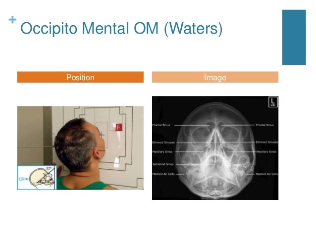 Source: slideshare.net
The side to the imaged should be positioned nearest to the table. This type of sinus can pose difficulties in interpreting radiological appearance. Is recommended for evaluation of paranasal sinuses. About press copyright contact us creators advertise developers terms privacy policy & safety how youtube works test new features press copyright contact us creators. For each setup in the tables, there is a picture demonstrating.
 Source: akehntu.blogspot.com
Position of patient lying on the side (left or right) with a vertical beam angled at 15 degrees. The side to the imaged should be positioned nearest to the table. The overlying petrosal bone masks the maxillary sinus.by the way, you can see the superior orbital fissure and the round foramen beautifully on this projection. Based on the acr appropriateness criteria ©, ct sinus. Position of part remove dentures, facial jewelry, earrings, and anything from the hair.
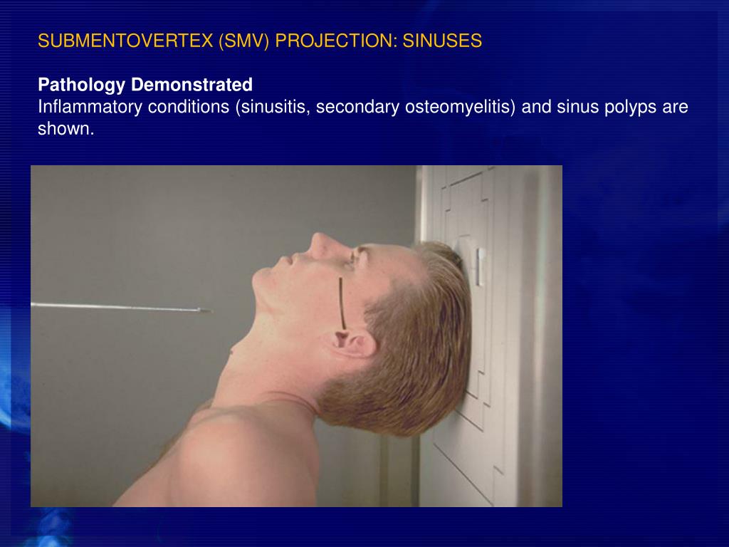 Source: slideserve.com
About press copyright contact us creators advertise developers terms privacy policy & safety how youtube works test new features press copyright contact us creators. Paranasal sinuses water’s view caldwell view lateral view submentovertical view right and left vertical view. Symptoms of other infection in the head area. Frontal sinuses projected above the frontonasal suture, anterior ethmoid air cells visualised lateral to each nasal bone directly below the frontal sinuses. Other sinuses related problems such as deviated septum.
 Source: pinterest.ph
An opaque sinus may be caused by the thickening of the bony walls, small asymmetric antra, or improper centering and rotation of the head. Sometimes additional stacking is applied, allowing to examine defects in more detail. Patient positioned same as waters view, but with mouth opened. Is recommended for evaluation of paranasal sinuses. Patient positioning for skull radiography.
 Source: pinterest.com
Symptoms of other infection in the head area. Patients can be imaged either erect or recumbent. The conventional paranasal sinus examination should consist of a minimum of three views: The patient’s head should be tilted by 15 degrees. The image depicts the frontal, ethmoidal, and sphenoid sinuses.
 Source: slideshare.net
This type of sinus can pose difficulties in interpreting radiological appearance. Position of patient lying on the side (left or right) with a vertical beam angled at 15 degrees. Patient positioning for skull radiography. Sinusitis, secondary osteomyelitis and sinus polyps. Position of part remove dentures, facial jewelry, earrings, and anything from the hair.
 Source: smartypance.com
Symptoms of other infection in the head area. Sometimes additional stacking is applied, allowing to examine defects in more detail. This view aids in visualizing the paranasal sinuses, especially the frontal sinus.it can help to assess 4 inflammatory conditions such as sinusitis and secondary osteomyelitis, and sinus polyps or cysts. Is recommended for evaluation of paranasal sinuses. About press copyright contact us creators advertise developers terms privacy policy & safety how youtube works test new features press copyright contact us creators.
 Source: youtube.com
Patient positioning for skull radiography. Radt 210 radiographic positioning iiisan diego mesa collegeradiographic positioning of the paranasal sinuses Patient positioning for skull radiography. This view must demonstrate sphenoid sinus through the open mouth (patient�s mouth is placed on table). This type of sinus can pose difficulties in interpreting radiological appearance.
 Source: humangrossanatomy.com
Sinusitis, secondary osteomyelitis and sinus polyps. The patient’s head should be tilted by 15 degrees. 3 views • waters • caldwell • lateral • all films should be done upright with horizontal beam. Position of part remove dentures, facial jewelry, earrings, and anything from the hair. This type of sinus can pose difficulties in interpreting radiological appearance.
If you find this site good, please support us by sharing this posts to your own social media accounts like Facebook, Instagram and so on or you can also bookmark this blog page with the title sinuses x ray positioning by using Ctrl + D for devices a laptop with a Windows operating system or Command + D for laptops with an Apple operating system. If you use a smartphone, you can also use the drawer menu of the browser you are using. Whether it’s a Windows, Mac, iOS or Android operating system, you will still be able to bookmark this website.


