Mitral valve prolapse mmode
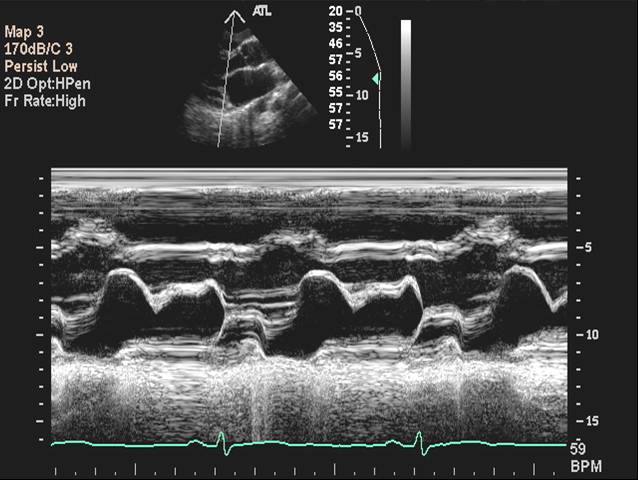
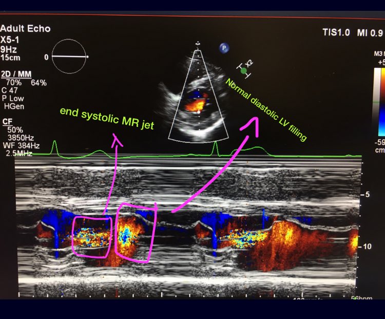
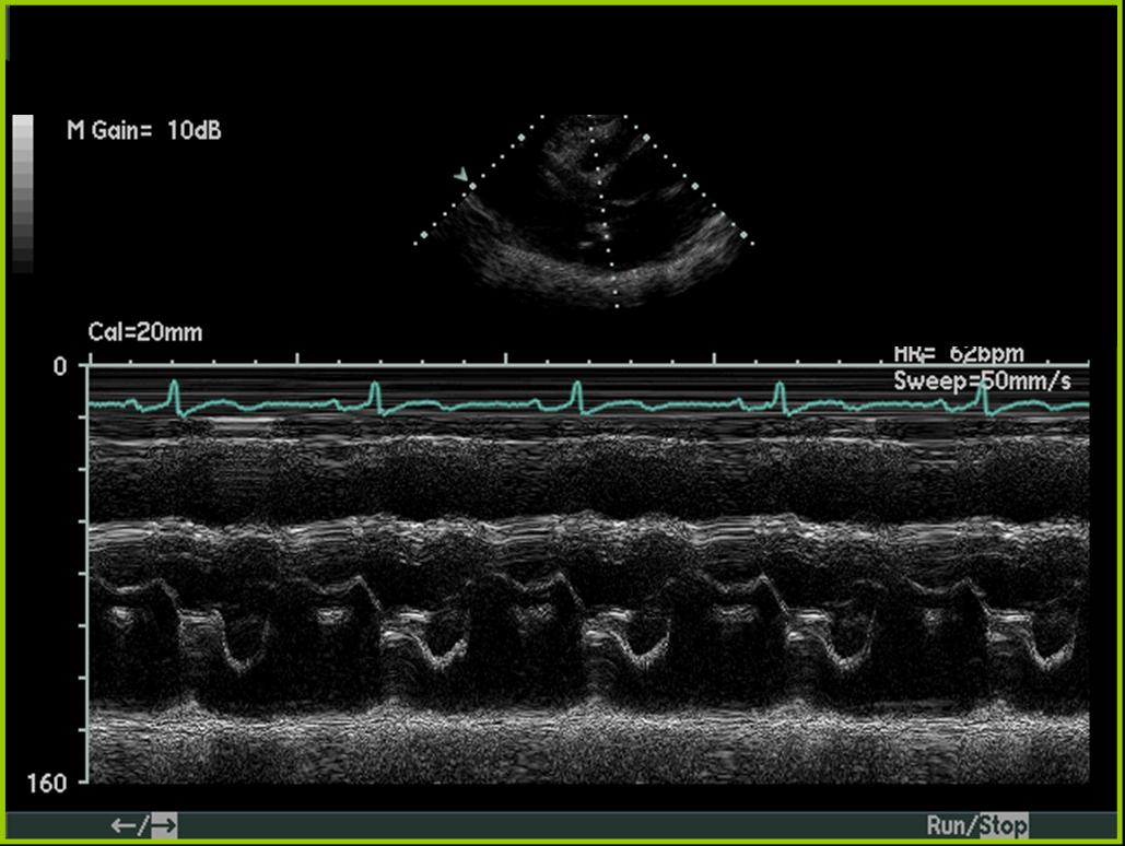
Mitral Valve Prolapse Mmode. While this is a single line of sight scans, a lot of information about the left ventricular function, mitral valve. In rare cases, it may present with sudden cardiac death, endocarditis or a stroke. 6.2 m mode in mitral valve prolapse; 6.4 m mode in mitral stenosis;
 Pin by nonas arc on Mitral Valve Prolapse (MVP) Mitral valve From pinterest.com
They bulge backward (prolapse) like a parachute into the heart�s left upper chamber as the heart squeezes (contracts). There were 46 men and 79 women; 6.1 m mode in aortic stenosis; It is a benign condition. Mvp is defined as single or bileaflet prolapse, with or without leaflet thickening. [1] [2] [3] the condition affects nearly 3% of the us population.
While this is a single line of sight scans, a lot of information about the left ventricular function, mitral valve.
Mitral valve prolapse (mvp) is bending backwards of the mitral valve as it closes when the left ventricle contracts. Criteria for mvp • > 3mm late systolic buckling • pan systolic hammocking 5mm or more. 6.1 m mode in aortic stenosis; Mitral valve (mv) prolapse (mvp) is the most common cause of primary mitral regurgitation (mr) in developed countries. 6 m mode pathologic states. It is a benign condition.
 Source: researchgate.net
• m mode useful when 2d echo is equivocal. • m mode useful when 2d echo is equivocal. The left parasternal axis view shows the left ventricle, mitral valve, and aortic valve. Their mean age was 42. Mitral valve prolapse (mvp) is the most common valvular disease with a prevalence of 2%.
 Source: wikidoc.org
They bulge backward (prolapse) like a parachute into the heart�s left upper chamber as the heart squeezes (contracts). 6.2 m mode in mitral valve prolapse; There were 46 men and 79 women; Left ventricle is the lower left chamber which pumps oxygenated blood to the whole body. Mvp is defined as single or bileaflet prolapse, with or without leaflet thickening.
 Source: twitter.com
Left ventricle is the lower left chamber which pumps oxygenated blood to the whole body. Mitral valve prolapse (mvp) is a valvular heart disease characterized by the displacement of an abnormally thickened mitral valve leaflet into the left atrium during systole. Mitral valve prolapse (mvp) is the most common valvular. It is the primary form of myxomatous degeneration of the valve. The left parasternal axis view shows the left ventricle, mitral valve, and aortic valve.
 Source: medison.ru
Mitral valve prolapse is a type of heart valve disease that affects the valve between the left heart chambers. Mitral valve prolapse (mvp) is a valvular heart disease characterized by the displacement of an abnormally thickened mitral valve leaflet into the left atrium during systole. It displays mitral valve motion and was mainly used to quantify mitral stenosis as well as visualize mitral valve prolapse and systolic anterior motion (sam) of. M mode • first echocardiographic technique to diagnose mvp. Because of the dependence of the ultrasound beam, however.
 Source: researchgate.net
Mitral valve prolapse (mvp) is bending backwards of the mitral valve as it closes when the left ventricle contracts. 6.1 m mode in aortic stenosis; What you need to know. Mitral valve prolapse (mvp) is a valvular heart disease characterized by the displacement of an abnormally thickened mitral valve leaflet into the left atrium during systole. Mvp is defined as single or bileaflet prolapse, with or without leaflet thickening.
 Source: wikidoc.org
While this is a single line of sight scans, a lot of information about the left ventricular function, mitral valve. 6.4 m mode in mitral stenosis; There are various types of mvp, broadly classified as classic and nonclassic. 6.3 m mode in hypertrophic cardiomyopathy; While mvp is primarily a disorder of the mv apparatus, few studies have suggested concomitant abnormalities in the left ventricle (lv).
 Source: medison.ru
6.4 m mode in mitral stenosis; M mode • first echocardiographic technique to diagnose mvp. Mitral valve (mv) prolapse (mvp) is the most common cause of primary mitral regurgitation (mr) in developed countries. The left parasternal axis view shows the left ventricle, mitral valve, and aortic valve. In rare cases, it may present with sudden cardiac death, endocarditis or a stroke.
 Source: researchgate.net
Because of the dependence of the ultrasound beam, however. 6 m mode pathologic states. It has generally a benign course; It displays mitral valve motion and was mainly used to quantify mitral stenosis as well as visualize mitral valve prolapse and systolic anterior motion (sam) of. In severe cases of classic mvp, complications include mitral.
 Source: researchgate.net
The left parasternal axis view shows the left ventricle, mitral valve, and aortic valve. In rare cases, it may present with sudden cardiac death, endocarditis or a stroke. 6.3 m mode in hypertrophic cardiomyopathy; 6.1 m mode in aortic stenosis; While this is a single line of sight scans, a lot of information about the left ventricular function, mitral valve.
 Source: wikidoc.org
Their mean age was 42. While mvp is primarily a disorder of the mv apparatus, few studies have suggested concomitant abnormalities in the left ventricle (lv). Their mean age was 42. It displays mitral valve motion and was mainly used to quantify mitral stenosis as well as visualize mitral valve prolapse and systolic anterior motion (sam) of. Celiac artery stenosis (cas) may be caused by atherosclerotic degeneration or compression exerted by the arched ligament of the diaphragm.
 Source: openi.nlm.nih.gov
Because of the dependence of the ultrasound beam, however. It is a benign condition. However, recent findings suggested an association between mvp and complex arrhythmias and eventually cardiac arrest and for this reason, it is also called arrhythmogenic mvp. Mitral valve prolapse (mvp) is bending backwards of the mitral valve as it closes when the left ventricle contracts. • m mode useful when 2d echo is equivocal.
 Source: pinterest.jp
6.2 m mode in mitral valve prolapse; It is the primary form of myxomatous degeneration of the valve. Mitral valve prolapse (mvp) is bending backwards of the mitral valve as it closes when the left ventricle contracts. However, recent findings suggested an association between mvp and complex arrhythmias and eventually cardiac arrest and for this reason, it is also called arrhythmogenic mvp. 6.1 m mode in aortic stenosis;
 Source: wikidoc.org
6.5 m mode in tamponade; There were 46 men and 79 women; In rare cases, it may present with sudden cardiac death, endocarditis or a stroke. [1] [2] [3] the condition affects nearly 3% of the us population. Their mean age was 42.
 Source: pinterest.com
There are various types of mvp, broadly classified as classic and nonclassic. However, recent findings suggested an association between mvp and complex arrhythmias and eventually cardiac arrest and for this reason, it is also called arrhythmogenic mvp. (please see companion dvd for corresponding video.) Mitral valve prolapse (mvp) is the most common valvular disease with a prevalence of 2%. M mode • first echocardiographic technique to diagnose mvp.
 Source: researchgate.net
Mitral valve prolapse (mvp) is the most common valvular. They bulge backward (prolapse) like a parachute into the heart�s left upper chamber as the heart squeezes (contracts). Their mean age was 42. Mitral valve (mv) prolapse (mvp) is the most common cause of primary mitral regurgitation (mr) in developed countries. It has generally a benign course;
 Source: sloanwebdesign.blogspot.com
Mitral valve is the valve between the left upper and lower chambers of the heart. 7 other m mode findings. However, recent findings suggested an association between mvp and complex arrhythmias and eventually cardiac arrest and for this reason, it is also called arrhythmogenic mvp. Subjects who experience this complication are in. While mvp is primarily a disorder of the mv apparatus, few studies have suggested concomitant abnormalities in the left ventricle (lv).
 Source: researchgate.net
Mitral valve prolapse is a type of heart valve disease that affects the valve between the left heart chambers. Mvp is defined as single or bileaflet prolapse, with or without leaflet thickening. M mode • first echocardiographic technique to diagnose mvp. There are various types of mvp, broadly classified as classic and nonclassic. It is the primary form of myxomatous degeneration of the valve.
 Source: medison.ru
• very specific, not sensitive. In severe cases of classic mvp, complications include mitral. In rare cases, it may present with sudden cardiac death, endocarditis or a stroke. 6.2 m mode in mitral valve prolapse; [1] [2] [3] the condition affects nearly 3% of the us population.
If you find this site value, please support us by sharing this posts to your preference social media accounts like Facebook, Instagram and so on or you can also save this blog page with the title mitral valve prolapse mmode by using Ctrl + D for devices a laptop with a Windows operating system or Command + D for laptops with an Apple operating system. If you use a smartphone, you can also use the drawer menu of the browser you are using. Whether it’s a Windows, Mac, iOS or Android operating system, you will still be able to bookmark this website.


