M mode mitral valve
M Mode Mitral Valve. 6.6 m mode aortic regurgitation; Choose parasternal long (left parasternal view.long axis) from the list <<positions>> or find the same position with 3d transducer. This technique was initially used primarily for the preoperative study of mitral valve stenosis. The velocity of propagation of flow (vp) from the lv base toward the apex is measured in early diastole.
 Mitral stenosis echocardiography wikidoc From wikidoc.org
Axial resolution allows for the vertical distance between the maximal early excursion of the anterior leaflet of the mitral valve and the interventricular septum. 6 m mode pathologic states. The velocity of propagation of flow (vp) from the lv base toward the apex is measured in early diastole. 6.4 m mode in mitral stenosis; 6.6 m mode aortic regurgitation; The upper panel shows the doming of anterior mitral leaflet in diastole.
Two dimensional echocardiography appears to be superior to m mode echocardiography in the diagnosis of a flail leaflet, papillary muscle dysfunction and cleft mitral valve.
(please see companion dvd for corresponding video.) 6.3 m mode in hypertrophic cardiomyopathy; Axial resolution allows for the vertical distance between the maximal early excursion of the anterior leaflet of the mitral valve and the interventricular septum. I guess it is a normal variant. 6.5 m mode in tamponade; The mitral valve is the first structure of the heart identified through echocardiogram.
 Source: researchgate.net
The exact answer is not known. Though it is not common , i have seen in few. Infrequently , m mode echo will record a triphasic pattern. 6.3 m mode in hypertrophic cardiomyopathy; The exact answer is not known.
 Source: wikidoc.org
6.2 m mode in mitral valve prolapse; Above is a picture of an echocardiogram with a. Choose parasternal long (left parasternal view.long axis) from the list <<positions>> or find the same position with 3d transducer. 6.4 m mode in mitral stenosis; When doppler echo was not available, m mode of the pulmonary valve was an important tool to assess pulmonary hypertension.
 Source: medison.ru
Because of the dependence of the ultrasound beam, however. Though it is not common , i have seen in few. (please see companion dvd for corresponding video.) 6.3 m mode in hypertrophic cardiomyopathy; Infrequently , m mode echo will record a triphasic pattern.
 Source: wikidoc.org
Both m mode and two dimensional echocardiography are useful in determining the origin of mitral regurgitation. 6 m mode pathologic states. Click the button <<m>> to move to one dimensional echocardiography ().; When doppler echo was not available, m mode of the pulmonary valve was an important tool to assess pulmonary hypertension. 6.5 m mode in tamponade;
 Source: wikidoc.org
6.6 m mode aortic regurgitation; 6.1 m mode in aortic stenosis; The upper panel shows the doming of anterior mitral leaflet in diastole. (please see companion dvd for corresponding video.) 6.4 m mode in mitral stenosis;
 Source: pinterest.com
Above is a picture of an echocardiogram with a. 6 m mode pathologic states. Click the button <<m>> to move to one dimensional echocardiography ().; Other m modes ©2020 by echocardiographer.org. 6.4 m mode in mitral stenosis;
 Source: researchgate.net
6.3 m mode in hypertrophic cardiomyopathy; The velocity of propagation of flow (vp) from the lv base toward the apex is measured in early diastole. Because of the dependence of the ultrasound beam, however. What about a little bit of theory before beginning? 6.1 m mode in aortic stenosis;
 Source: medison.ru
The exact answer is not known. The mitral valve is the first structure of the heart identified through echocardiogram. Inge edler and physicist hellmuth hertz marked the beginning of a new noninvasive diagnostic technique. Because of the dependence of the ultrasound beam, however. 6 m mode pathologic states.
 Source: researchgate.net
6.6 m mode aortic regurgitation; 6 m mode pathologic states. The exact answer is not known. Because of the dependence of the ultrasound beam, however. Two dimensional echocardiography appears to be superior to m mode echocardiography in the diagnosis of a flail leaflet, papillary muscle dysfunction and cleft mitral valve.
 Source: researchgate.net
Run the echocardiography online simulator; Click the button <<m>> to move to one dimensional echocardiography ().; The velocity of propagation of flow (vp) from the lv base toward the apex is measured in early diastole. 17 to 30 mm 21. Run the echocardiography online simulator;
 Source: researchgate.net
Choose parasternal long (left parasternal view.long axis) from the list <<positions>> or find the same position with 3d transducer. 6.6 m mode aortic regurgitation; The mitral valve is the first structure of the heart identified through echocardiogram. Choose parasternal long (left parasternal view.long axis) from the list <<positions>> or find the same position with 3d transducer. Infrequently , m mode echo will record a triphasic pattern.
 Source: heart-valve-surgery.com
Click the button <<m>> to move to one dimensional echocardiography ().; 6.6 m mode aortic regurgitation; If these three techniques are contraindicated, a transesophageal echocardiogram can be performed. (please see companion dvd for corresponding video.) 6 m mode pathologic states.
 Source: medison.ru
I guess it is a normal variant. Two dimensional echocardiography appears to be superior to m mode echocardiography in the diagnosis of a flail leaflet, papillary muscle dysfunction and cleft mitral valve. 6.4 m mode in mitral stenosis; 6.5 m mode in tamponade; 7 other m mode findings.
 Source: sites.austincc.edu
This technique was initially used primarily for the preoperative study of mitral valve stenosis. Both m mode and two dimensional echocardiography are useful in determining the origin of mitral regurgitation. 6.5 m mode in tamponade; The velocity of propagation of flow (vp) from the lv base toward the apex is measured in early diastole. Other m modes ©2020 by echocardiographer.org.
 Source: wikidoc.org
Though it is not common , i have seen in few. 7 other m mode findings. Two dimensional echocardiography appears to be superior to m mode echocardiography in the diagnosis of a flail leaflet, papillary muscle dysfunction and cleft mitral valve. Click the button <<m>> to move to one dimensional echocardiography ().; 6 m mode pathologic states.
 Source: wikidoc.org
The mitral valve is the first structure of the heart identified through echocardiogram. 6.3 m mode in hypertrophic cardiomyopathy; The velocity of propagation of flow (vp) from the lv base toward the apex is measured in early diastole. The upper panel shows the doming of anterior mitral leaflet in diastole. Though it is not common , i have seen in few.
 Source: researchgate.net
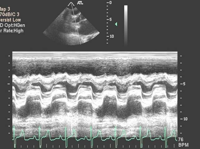
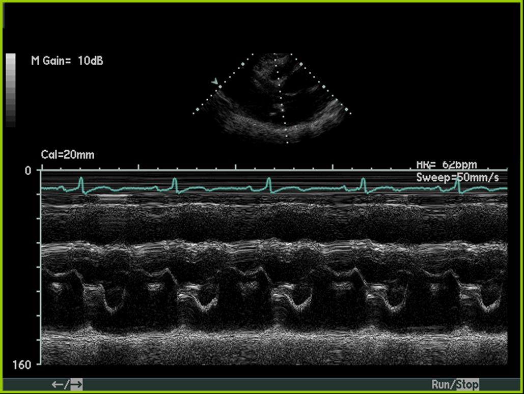
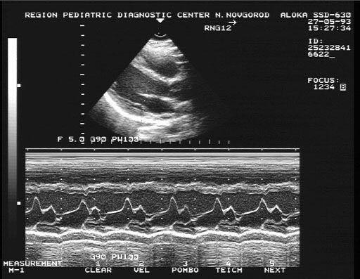
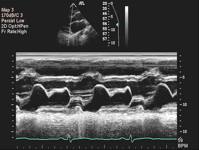
Anterior mitral leaflet has a classical m shaped motion. 6 m mode pathologic states. If these three techniques are contraindicated, a transesophageal echocardiogram can be performed. 17 to 30 mm 21. The velocity of propagation of flow (vp) from the lv base toward the apex is measured in early diastole.
 Source: researchgate.net
(please see companion dvd for corresponding video.) 6.2 m mode in mitral valve prolapse; Inge edler and physicist hellmuth hertz marked the beginning of a new noninvasive diagnostic technique. The upper panel shows the doming of anterior mitral leaflet in diastole. Both m mode and two dimensional echocardiography are useful in determining the origin of mitral regurgitation.
If you find this site beneficial, please support us by sharing this posts to your own social media accounts like Facebook, Instagram and so on or you can also save this blog page with the title m mode mitral valve by using Ctrl + D for devices a laptop with a Windows operating system or Command + D for laptops with an Apple operating system. If you use a smartphone, you can also use the drawer menu of the browser you are using. Whether it’s a Windows, Mac, iOS or Android operating system, you will still be able to bookmark this website.


