Echogenic foci in gallbladder
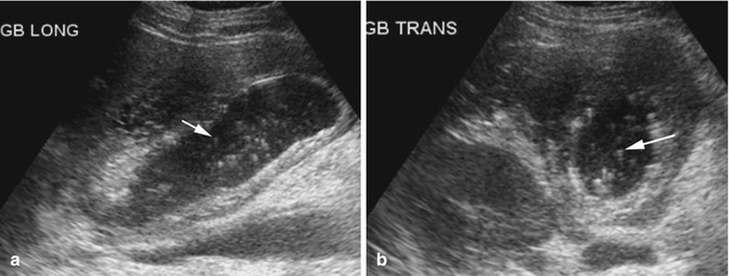
Echogenic Foci In Gallbladder. In most cases, gallbladder sludge does not. Abdominal sonography in 7 patients with acute biliary disease revealed focal echo collections within the wall of the gallbladder in addition to cholelithiasis and diffuse mural thickening. Obstetric sonograms of 26 fetuses with echogenic material in the gallbladder were reviewed to describe the sonographic findings and clinical significance. Gallbladder is partially distended, showing two echogenic foci , not casting posterior acoustic shadowing , measuring 3.0 mm.
 Imaging of Acute Abdominal Pain in the Pediatric Population From healio.com
Although fatty liver disease may progress, it can also be reversed with diet and lifestyle changes. In 3 patients, microscopy of the hyperchoic tissue showed. The majority are cholesterol polyps. Echogenic material in the gallbladder is debris formed in the bile of the gallbladder. Drinking beet juice or eating beets. Irfan tariq, md answered this.
Obstetric sonograms of 26 fetuses with echogenic material in the gallbladder were reviewed to describe the sonographic findings and clinical significance.
Abdominal sonography in 7 patients with acute biliary disease revealed focal echo collections within the wall of the gallbladder in addition to cholelithiasis and diffuse mural thickening. Gallbladder, liver, pancreas & spleen issues. Foci of increased acoustic density in the thickness of the myometrium are observed in women with diabetes mellitus during the menopause or after scraping. Obstetric sonograms of 26 fetuses with echogenic material in the gallbladder were reviewed to describe the sonographic findings and clinical significance. It is a relatively common and benign cause of diffuse or focal gallbladder wall thickening, most easily seen on ultrasound and mri. A segment of an organ or tissue with a high density for ultrasonic waves is a hyperechoic formation.
 Source: radiologykey.com
Irfan tariq, md answered this. Also known as gallbladder sludge, it shifts about from time to time within the gallbladder. In the gallbladder, such a seal may indicate: A high content of fat in the liver is indicative of fatty liver disease. Gallbladder polyps are relatively frequent, seen in up to 9% of the population 1,7,12,14.
 Source: researchgate.net
It is a relatively common and benign cause of diffuse or focal gallbladder wall thickening, most easily seen on ultrasound and mri. It shows multiple small echogenic foci some with posterior shadowing and some has no posterior shadowing in the gallbladder. Along with trying home remedies. These echoes do not cast an acoustic shadow. Gallbladder is partially distended, showing two echogenic foci , not casting posterior acoustic shadowing , measuring 3.0 mm.
 Source: semanticscholar.org
Echogenic foci in the gallbladder are usually small polyps. Drinking unrefined olive oil on an empty stomach. Hyperechoic formation in the gallbladder. If you have a fever. They can be seen on an ultrasound and are often benign.
 Source: clinicalultrasound.org
Also known as gallbladder sludge, it shifts about from time to time within the gallbladder. Along with trying home remedies. Foci of increased acoustic density in the thickness of the myometrium are observed in women with diabetes mellitus during the menopause or after scraping. The report sounds like gallstone (echogenic focus and sludge) with some irritation of the gallbladder (wall thickening). Drinking pear juice or eating pears.
 Source: researchgate.net
Also known as gallbladder sludge, it shifts about from time to time within the gallbladder. Abdominal sonography in 7 patients with acute biliary disease revealed focal echo collections within the wall of the gallbladder in addition to cholelithiasis and diffuse mural thickening. An echogenic liver is an ultrasound reading that indicates a higher level of fat in the liver. The majority are cholesterol polyps. Gallbladder, liver, pancreas & spleen issues.
 Source: criticalcare-sonography.com
There is a 0.5 cm nonshadowing echogenic focus along the wall the fundus of the gallbladder that is nonmobile. It shows multiple small echogenic foci some with posterior shadowing and some has no posterior shadowing in the gallbladder. They can be seen on an ultrasound and are often benign. A segment of an organ or tissue with a high density for ultrasonic waves is a hyperechoic formation. A prospective study was conducted, comparing in vivo and in vitro sonography of the gallbladder with histopathological findings.
 Source: radiology-information.blogspot.com
An adenomatous polyp is less likely however followup is recommended to ensure stability. It shows multiple small echogenic foci some with posterior shadowing and some has no posterior shadowing in the gallbladder. In most cases, gallbladder sludge does not. Along with trying home remedies. If you have a fever.
 Source: radiologykey.com
The report sounds like gallstone (echogenic focus and sludge) with some irritation of the gallbladder (wall thickening). It is a relatively common and benign cause of diffuse or focal gallbladder wall thickening, most easily seen on ultrasound and mri. The report sounds like gallstone (echogenic focus and sludge) with some irritation of the gallbladder (wall thickening). In 3 patients, microscopy of the hyperchoic tissue showed. The majority are cholesterol polyps.
 Source: researchgate.net
Also known as gallbladder sludge, it shifts about from time to time within the gallbladder. This likely represents a gallbladder polyp. Along with trying home remedies. A prospective study was conducted, comparing in vivo and in vitro sonography of the gallbladder with histopathological findings. Gallbladder polyps are relatively frequent, seen in up to 9% of the population 1,7,12,14.
 Source: radiopaedia.org
Along with trying home remedies. The layering echogenic calculi produce posterior acoustic shadowing as marked. An echogenic liver is an ultrasound reading that indicates a higher level of fat in the liver. Asymptomatic gallbladder polyps do not seem to raise the risk of gallbladder cancer 19. Foci of increased acoustic density in the thickness of the myometrium are observed in women with diabetes mellitus during the menopause or after scraping.
 Source: bestkidneyfailure.blogspot.com
Applying hot water packs externally. The layering echogenic calculi produce posterior acoustic shadowing as marked. Drinking unrefined olive oil on an empty stomach. Echogenic foci in the gallbladder are usually small polyps. Applying hot water packs externally.
 Source: radiopaedia.org
Also known as gallbladder sludge, it shifts about from time to time within the gallbladder. Echogenic foci in the gallbladder are usually small polyps. When striving to protect your liver, aim to drink lots of water, eat high. A prospective study was conducted, comparing in vivo and in vitro sonography of the gallbladder with histopathological findings. Asymptomatic gallbladder polyps do not seem to raise the risk of gallbladder cancer 19.
 Source: clinicalultrasound.org
Drinking pear juice or eating pears. The pathognomonic finding of adenomyomatosis is the presence of hyperechoic foci, most commonly noted in the anterior wall of the gallbladder, which produces a comet tail artifact that decreases in width and amplitude more posteriorly ( fig. They can be seen on an ultrasound and are often benign. This likely represents a gallbladder polyp. The layering echogenic calculi produce posterior acoustic shadowing as marked.
 Source: researchgate.net
When striving to protect your liver, aim to drink lots of water, eat high. Drinking pear juice or eating pears. Gestational age at the time of diagnosis ranged from 28 to 42 weeks (mean, 36.2 weeks). Drinking unrefined olive oil on an empty stomach. If you have a fever.
 Source: researchgate.net
Gallbladder polyps are relatively frequent, seen in up to 9% of the population 1,7,12,14. Applying hot water packs externally. Irfan tariq, md answered this. The majority are cholesterol polyps. The layering echogenic calculi produce posterior acoustic shadowing as marked.
 Source: onlinejets.org
Drinking pear juice or eating pears. Gallbladder, liver, pancreas & spleen issues. The layering echogenic calculi produce posterior acoustic shadowing as marked. Although fatty liver disease may progress, it can also be reversed with diet and lifestyle changes. Along with trying home remedies.
 Source: todaysveterinarypractice.com
Asymptomatic gallbladder polyps do not seem to raise the risk of gallbladder cancer 19. In the gallbladder, such a seal may indicate: The majority are cholesterol polyps. It shows multiple small echogenic foci some with posterior shadowing and some has no posterior shadowing in the gallbladder. The layering echogenic calculi produce posterior acoustic shadowing as marked.
 Source: healio.com
Statistically this represents a cholesterol polyp. A high content of fat in the liver is indicative of fatty liver disease. They can be seen on an ultrasound and are often benign. Applying hot water packs externally. If you have a fever.
If you find this site helpful, please support us by sharing this posts to your preference social media accounts like Facebook, Instagram and so on or you can also bookmark this blog page with the title echogenic foci in gallbladder by using Ctrl + D for devices a laptop with a Windows operating system or Command + D for laptops with an Apple operating system. If you use a smartphone, you can also use the drawer menu of the browser you are using. Whether it’s a Windows, Mac, iOS or Android operating system, you will still be able to bookmark this website.


