Coccyx x ray positioning
Coccyx X Ray Positioning. Palpate bottom of coccyx to center 4. Ensure no rotation of the pelvis. • patient position—supine position • part position • align midsagittal plane to cr and midline of table and/or ir. The patient lies on his back, bends his legs at the knee and hip joints (or only at the knees).
 Coccyx Fracture From pt.slideshare.net
The arms are extended along the body. #sacrum#coccyxhello!!!!what’s up guys, kaise ho dosto? Cr angled caudad 10 °. Ensure no rotation of body and pelvis for true lateral position. 10 degree caudal tube angle. Sacrum • pathology of the sacrum, including fracture.
10 degree caudal tube angle.
#sacrum#coccyxhello!!!!what’s up guys, kaise ho dosto? Ensure pelvis and torso are in a true lateral position; The patient is in a lateral recumbent position 1. Legs extended with a support under the knees. In coccygodynia, pain is most severe in the sitting position. It helps to visualize pathology of the sacrum and coccyx, and investigates the cause of sacral and c occyx pain in both acute and chronic conditions.
 Source: coccygectomy.org
• patient position—supine position • part position • align midsagittal plane to cr and midline of table and/or ir. Coccygeal segments should appear open on radiograph, if not they may be fused, or an increase on central ray angulation. Patient in the lateral recumbent position; • patient position—supine position • part position • align midsagittal plane to cr and midline of table and/or ir. Ensure no rotation of body and pelvis for true lateral position.
 Source: slideserve.com
Aur samaj lenge ki sacr. This can dramatically decrease the amount of radiation delivered to the patient and can especially decrease the amount of radiation delivered to. Cr angled caudad 10 °. Align long axis of sacrum and coccyx to central ray and to midline of table or gird. Founder and director at tailbone pain center.
 Source: isjonline.com
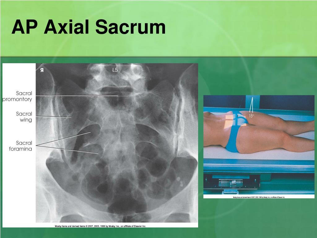
In coccygodynia, pain is most severe in the sitting position. Cr angled caudad 10 °. Aur samaj lenge ki sacr. The arms are extended along the body. 3 views • ap sacrum with central ray angled 15 degrees cephalad • ap coccyx with central ray angled 10 degrees caudad • lateral sacrum/coccyx.
 Source: lumen.luc.edu
This observation has been common to all roentgenologists for many years and does not. Center midsagittal and 2 above symphysis pubis 4. Align long axis of sacrum and coccyx to central ray and to midline of table or gird. The patient is in a lateral recumbent position 1. It shows which joint is dislocating, allowing the doctor to give a corticosteroid injection in the right place.

The coccyx lateral view is used to show the most distal region of the spine in a lateral position. This can dramatically decrease the amount of radiation delivered to the patient and can especially decrease the amount of radiation delivered to. Correct coccyx and central ray alignment demonstrate coccyx free of superimposition and projected superior to pubis. This observation has been common to all roentgenologists for many years and does not. In coccygodynia, pain is most severe in the sitting position.
 Source: pt.slideshare.net
8 x 10 film 2. Foye is an expert at treating tailbone pain (coccyx pain). 3 views • ap sacrum with central ray angled 15 degrees cephalad • ap coccyx with central ray angled 10 degrees caudad • lateral sacrum/coccyx. It is used in conjunction with the ap projection. Coccyx should appear equal distant from.
 Source: researchgate.net
3 views • ap sacrum with central ray angled 15 degrees cephalad • ap coccyx with central ray angled 10 degrees caudad • lateral sacrum/coccyx. In coccygodynia, pain is most severe in the sitting position. Legs extended with a support under the knees. The patient can be either on the left or right lateral recumbent position, depending on which is more comfortable; This can dramatically decrease the amount of radiation delivered to the patient and can especially decrease the amount of radiation delivered to.
 Source: coccyxpain.blogspot.com
Foye is an expert at treating tailbone pain (coccyx pain). The patient lies on his back, bends his legs at the knee and hip joints (or only at the knees). Ensure no rotation of body and pelvis for true lateral position. Patient supine on the table. The coccyx lateral view is used to show the most distal region of the spine in a lateral position.
 Source: pinterest.co.uk
The coccyx lateral view is used to show the most distal region of the spine in a lateral position. It is used to show sacrum and coccyx anatomy, and to investigate the cause of sacral and coccyx pain in both acute and chronic conditions. Align long axis of coccyx to cr and to midline of table/grid; Place a support under waist and between knees and ankkles to maintain patient position and ensure comfort. Cushion for patients head ;
 Source: viu-wit.blogspot.com
Sacrum • pathology of the sacrum, including fracture. This observation has been common to all roentgenologists for many years and does not. Founder and director at tailbone pain center. Ensure pelvis and torso are in a true lateral position; Patient supine on the table.

Sacrum • pathology of the sacrum, including fracture. Palpate bottom of coccyx to center 4. It is used in conjunction with the ap projection. This projection helps to visualize pathology of the coccyx, especially fractures.to minimize superimposition of structures over the coccyx region, the urinary bladder and large colon should ideally be emptied before this examination 1. Coccygeal segments should appear open on radiograph, if not they may be fused, or an increase on central ray angulation.
 Source: reddit.com
It is used to show sacrum and coccyx anatomy, and to investigate the cause of sacral and coccyx pain in both acute and chronic conditions. Dosto aj sacrum and coccyx ka. 10 degree caudal tube angle. The arms are extended along the body. #sacrum#coccyxhello!!!!what’s up guys, kaise ho dosto?
 Source: pinterest.ch
Tailbone pain is usually most painful while you are sitting, since sitting puts some of your body weight onto the tailbone (coccyx). The coccyx lateral view is used to show the most distal region of the spine in a lateral position. Legs extended with a support under the knees. Tap card to see definition. The arms are extended along the body.
 Source: viu-wit.blogspot.com
It helps to visualize pathology of the sacrum and coccyx, and investigates the cause of sacral and c occyx pain in both acute and chronic conditions. Radiographic views of sacrum and coccyx chandan prasad rajbhar tutor college of paramedical sciences tmu, moradabad. Dosto aj sacrum and coccyx ka positioning aur practical hum shabbi dekh lenge. Foye is an expert at treating tailbone pain (coccyx pain). 10 degree caudal tube angle.
 Source: samarpanphysioclinic.com
It helps to visualize pathology of the sacrum and coccyx, and investigates the cause of sacral and c occyx pain in both acute and chronic conditions. Sacrum • pathology of the sacrum, including fracture. It is used to show sacrum and coccyx anatomy, and to investigate the cause of sacral and coccyx pain in both acute and chronic conditions. It is used in conjunction with the ap projection. Foye is an expert at treating tailbone pain (coccyx pain).
 Source: gudangmedis.blogspot.com
Founder and director at tailbone pain center. Correct coccyx and central ray alignment demonstrate coccyx free of superimposition and projected superior to pubis. Palpate bottom of coccyx to center 4. The coccyx lateral view is used to show the most distal region of the spine in a lateral position. In coccygodynia, pain is most severe in the sitting position.
 Source: easynotecards.com
#sacrum#coccyxhello!!!!what’s up guys, kaise ho dosto? This can dramatically decrease the amount of radiation delivered to the patient and can especially decrease the amount of radiation delivered to. Align long axis of coccyx to cr and to midline of table/grid; Dosto aj sacrum and coccyx ka. It is used in conjunction with the ap projection.
 Source: radiopaedia.org
Ensure no rotation of body and pelvis for true lateral position. This prompted a study comparing lateral roentgenograms of the coccyx taken with the patient lying on the side. The coccyx lateral view is used to show the most distal region of the spine in a lateral position. • patient position—supine position • part position • align midsagittal plane to cr and midline of table and/or ir. This can dramatically decrease the amount of radiation delivered to the patient and can especially decrease the amount of radiation delivered to.
If you find this site adventageous, please support us by sharing this posts to your preference social media accounts like Facebook, Instagram and so on or you can also bookmark this blog page with the title coccyx x ray positioning by using Ctrl + D for devices a laptop with a Windows operating system or Command + D for laptops with an Apple operating system. If you use a smartphone, you can also use the drawer menu of the browser you are using. Whether it’s a Windows, Mac, iOS or Android operating system, you will still be able to bookmark this website.


